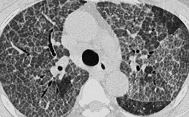
tree in bud radiology assistant
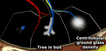
Tree-in-bud describes the appearance of an irregular and. Tree-in-bud TIB is a radiologic pattern seen on high-resolution chest CT reflecting bronchiolar mucoid impaction occasionally with additional involvement of adjacent alveoli.
Tree in bud radiology assistant.
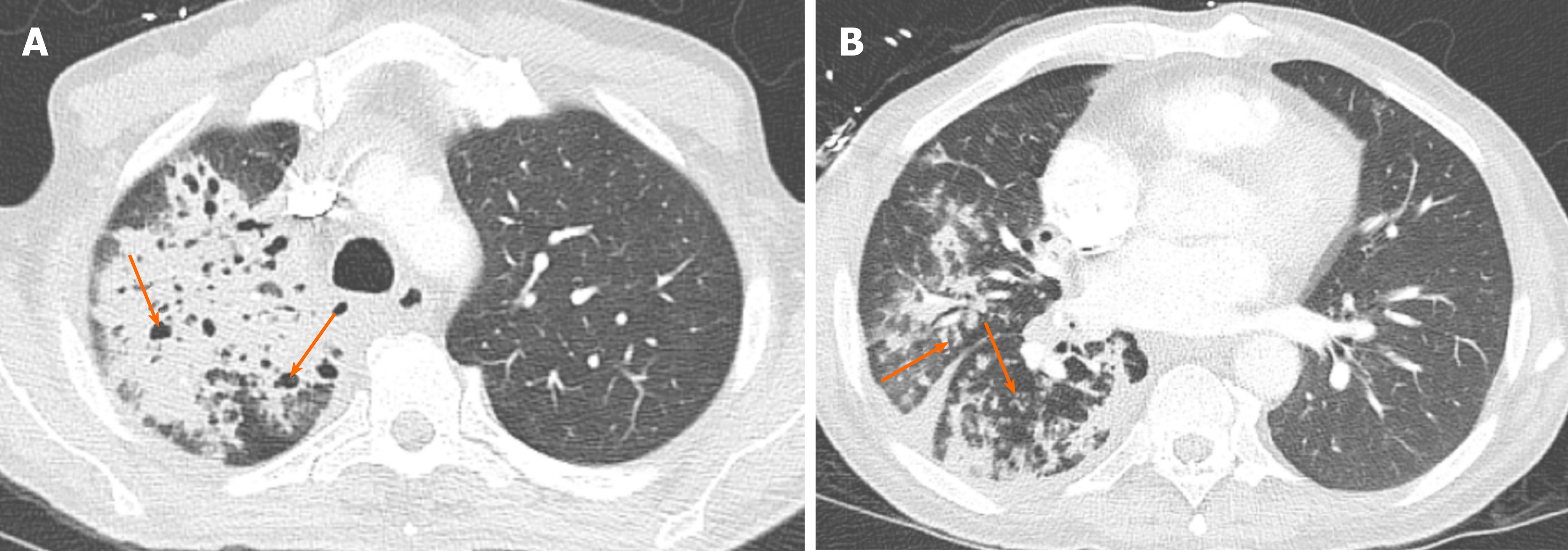
. The tree-in-bud pattern is commonly seen at thin-section computed tomography CT of the lungs. It represents dilated and impacted. Ad Indeed has a wide range of radiology Assistant jobs available.
Originally and still often thought to be specific to endobronchial Tb the sign is actually non-specific and is the. Ad Our Experts Provide 247 On-Call Support for Your Radiology Assignment. Get Better Pay A Flexible Schedule Fewer Admin Hassles.
Normal lobular bronchioles 1 mm in. Endobronchial spread of infection TB MAC any bacterial bronchopneumonia Airway disease associated with infection. The list of the most frequent differential diagnoses for tree-in-bud sign includes infections with Mycobacterium tuberculosis.
Friday April 8 2022. Tree-in-bud appearance represents dilated and fluid-filled ie. Tree-in-bud almost always indicates the presence of.
Tree-in-bud describes the appearance of an irregular and often nodular branching structure most easily identified in the lung periphery. In centrilobular nodules the recognition of tree-in-bud is of value for narrowing the differential diagnosis. The tree-in-bud sign is a common finding in HRCT scans.
Ad Its Time To Simplify Your Radiology Job Search. It consists of small centrilobular nodules of soft-tissue attenuation. Abnormal tree-in-bud bronchioles can be.
Areas of consolidation along with ground glass opacity involving the lingual contiguous with the inferior lateral portion of the left upper lobe abutting the left major fissure. Tree-in-bud describes the appearance of an irregular and often nodular branching structure most easily identified in the lung periphery. Indeed is looking for Radiologists Assistant to join our team.
Pus mucus or inflammatory exudate centrilobular bronchioles. Fig 7 Tree In Bud Sign Chest Ct Shows Tree In Bud Images Schematic Drawings And Corresponding Picture Refe Radiology Radiology Imaging Medical Radiography.

Chronic Airspace Disease Review Of The Causes And Key Computed Tomography Findings

Tree In Bud Sign Lung Radiology Reference Article Radiopaedia Org
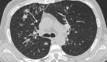
The Radiology Assistant Hrct Common Diagnoses

The Radiology Assistant Hrct Common Diagnoses

Common Examples Of Chest X Ray Consolidation Interstitial Nodule Mass And Atelectasis Medical Imaging Respiratory Therapy Radiology Student
The Radiology Assistant Hrct Basic Interpretation
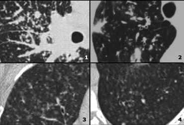
The Radiology Assistant Hrct Basic Interpretation
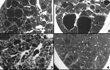
The Radiology Assistant Hrct Basic Interpretation
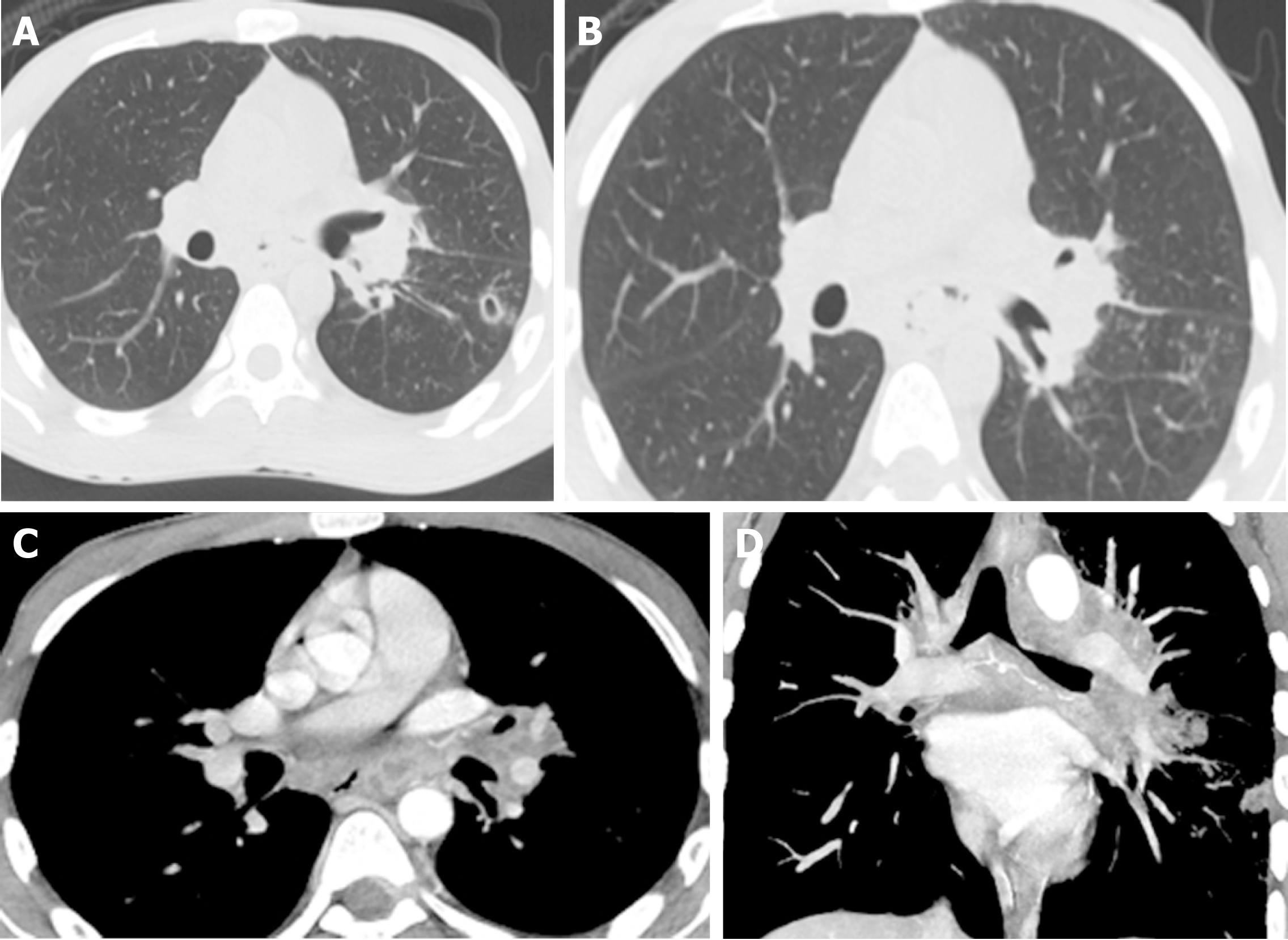
Tuberculous Esophagomediastinal Fistula With Concomitant Mediastinal Bronchial Artery Aneurysm Acute Upper Gastrointestinal Bleeding A Case Report

Cavity Consolidation With Multiple Areas Of Nodular Opacity Showing Tree In Bud Appearance Most Likely Possibility Of E Opacity Abstract Artwork Abstract
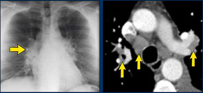
The Radiology Assistant Hrct Common Diagnoses

The Radiology Assistant Basic Interpretation Idiopathic Pulmonary Fibrosis Pulmonary Fibrosis Medical Radiography

Covid 19 Pneumonia Presenting With Multiple Nodules Mimicking Metastases An Atypical Case
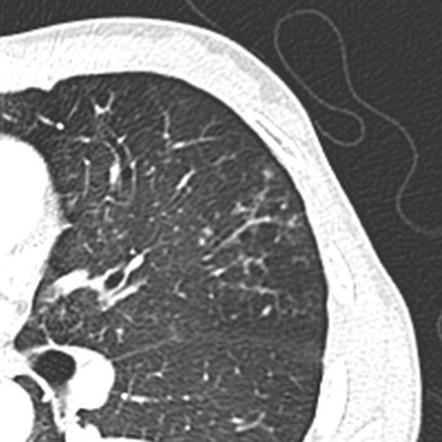
Tree In Bud Sign Lung Radiology Reference Article Radiopaedia Org


